-
- Autorefraktor / Keratometer
- Corneale modulare Systeme
- Farbtest & Farbtafeln
- Gläserkästen
- Handstücke
- Kontaktgläser Einweg und Mehrweg
- Kopfophthalmoskope
- Mesotest (Mesopisches Kontrastsehen)
- Messbrille / Probierbrille
- Nahsehtest
- Ophthalmometer
- Ophthalmoskopie direkt & indirekt
- Probiergläserkästen
- Refraktionsgläser
- Refraktionsprüfsätze / Brillenkästen
- Refraktionszubehör
- Refraktometer
- Scheitelbrechwertmesser
- Sehprüfgeräte
- Sehtafeln
- Sehzeichenprojektoren
- Spaltlampen & Zubehör
- Straßenverkehrsbezogener Sehtest
- Teststreifen
- Tonometer / Tonometrie
- Tonometerzubehör
-
- Abberometer und Wellenfrontabberometer
- Anomaloskop
- Elektrophysiologie
- Endothelmikroskope
- Funduskameras
- Hornhauttopographie
- IOL-Master
- Keratograph
- Keratometer
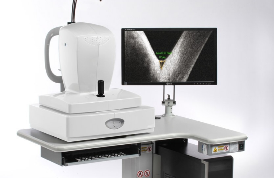
- Optische Kohärenztomographie (OCT)
- Pachymetrie
- Perimetrie und Zubehör
- Pupillometer
- Retina-Untersuchungsgeräte
- Retinometer
- Scheimpflugkameras
- Topographie (-Systeme)
- Ultraschall-Biometrie-Geräte
-
-
-
- Autoklaven & Sterilisationszubehör
- Chirurgielupen
- Crosslinking
- Crosslinking Zubehör
- Diamantmesser
- Glaukomchirurgie
- Hornhaut
- Hornhautkennzeichnung
- Hornhautstanzen
- Implantationsbesteck
- Injektoren
- Instrumente
- Instrumente (wiederverwendbar)
- Kataraktchirurgie
- Koagulationsgeräte
- Kryochirurgie-Geräte
- Lidsperrer
- Messer
- Mikrokeratome
- OP-Lampen
- OP-Liegen
- OP-Lüftungs- und Klimageräte
- OP-Mikroskope
- OP-Tische
- OP-Zubehör
- Operateurstühle
- Pendelmarkierer
- Phako-Zubehör
- Phakoemulsifikations-Geräte
- Pinzette
- Strabismusscheren
- Trepane
- Ultraschall-Reinigungsgeräte
- Vitrektomiemaschinen
- Vitrektomiezubehör
- Wetlab-.Zubehör
-
-
- Dienstleistungen
- Drehstühle & Drehhocker
- Ersatzlampen
- Fußschalter
- Gerätetische
- Glasschleifgeräte
- Karteischränke
- Lagerungshilfen
- Lupen Einweg und Mehrweg
- Patientenstühle
- Phoroptoren
- Refraktions- und Untersuchungseinheiten
- Refraktions- und Untersuchungsstühle
- Schreibtische
- Untersuchungseinheiten Umrüstsatz
- Untersuchungsgeräte
- Video-Betrachtungs und -Dokumentationseinrichtungen
-
- Anfärbelösungen
- Augenspülung
- Fluoreszenzangiographie
- Glaukom Implantate
- Hyaluronsäure
- Hygiene Produkte
- Implantate
- Instrumente (einmal)
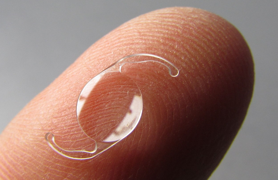
- Intraokularlinsen
- Iris Implantate
- Irisdiaphragma
- Kapselspannringe
- Nahtmaterial
- OP-Abdeckung
- OP-Bedarf
- OP-Mantel
- OP-Sets
- Ophthalmologische Gase
- Punctum Plugs
- Retina-Implantat
- Silikonöl
- Spüllösungen (intraokulare)
- Verbandslinsen
- Viskoelastika
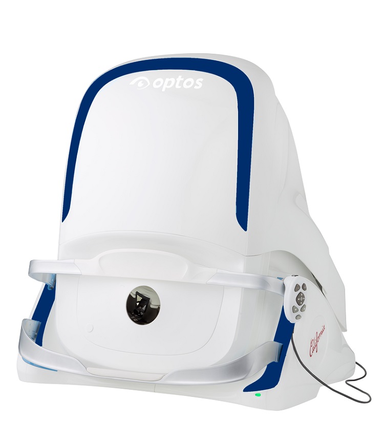

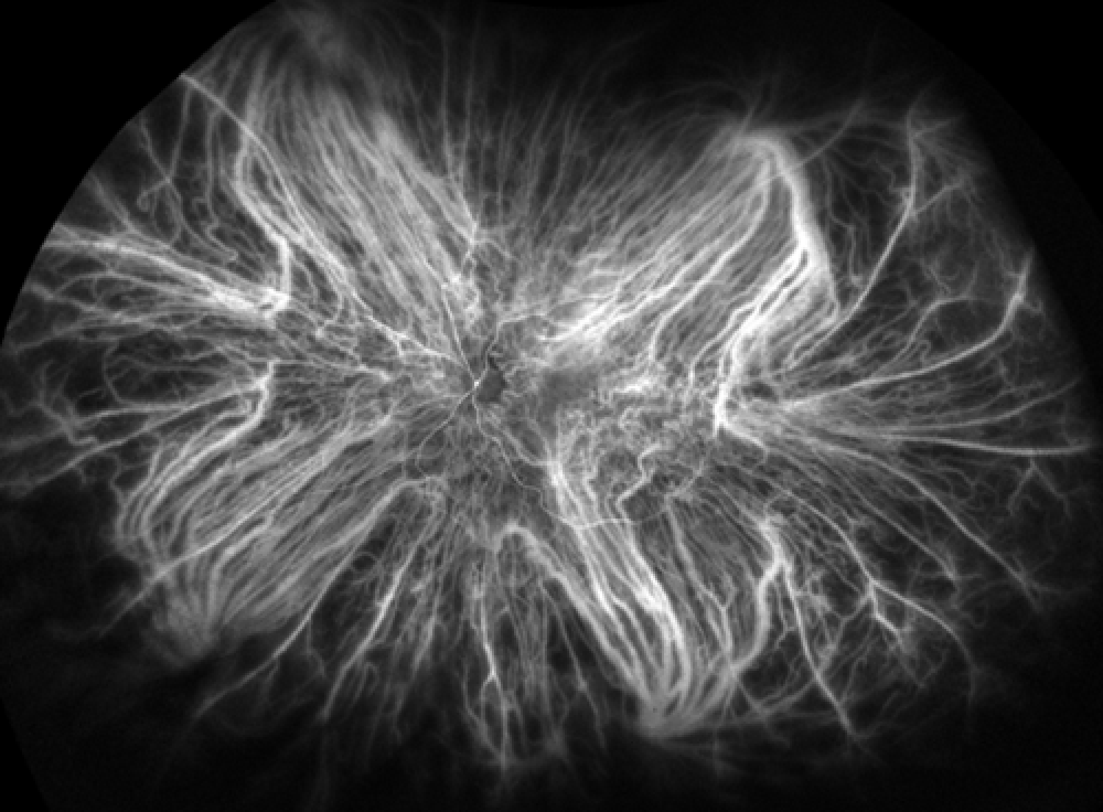
California - Optos Ultra-Weitwinkel (UWF™) Bildgebung der Netzhaut mit Angiographie-Optionen
Die innovative Optik von California liefert hochaufgelöste Bilder, die feine Details zeigen – unabhängig davon, ob die gesamte Netzhaut oder bestimmte Bereiche wie die Makula, der Sehnervenkopf oder kleine Pathologien vergrößert dargestellt werden.
Weitere ProduktinformationenCalifornia - Optos Ultra-Weitwinkel (UWF™) Bildgebung der Netzhaut mit Angiographie-Optionen
Ultra Wide Angle (UWF™) Imaging Device California
California is available in three models with six image modalities. In this way, the right model can be chosen depending on the requirements and budget of the practice:
•Color RG
•Color RGB
• Sensory retina (red-free)
• Choroidal
• Autofluorescence (af)
• Fluorescein Angiography (fa)
• ICG Angiography (icg)
optomap images are displayed in a uniform geometry that accurately depicts anatomical features of the retina. The automatic
Image registration enables pixel-by-pixel comparisons of images of different image modalities as well as from visit to visit.
Features and Benefits:
• Non-mydriatic retinal imaging in less than 1/2 second saves time and helps improve practice workflows.
• cSLO technology allows images to be taken through small pupils (2 mm) and often through cataracts.
• 3-in-1 Colour Depth Imaging™ Depth imaging provides critical clinical data from the retinal surface to the choroid.
• Green laser autofluorescence minimizes patient exposure to blue light and provides more detailed images of the macula and optic nerve head.
• The ability to overlay images makes it easier to compare images from different image modes as well as different post-observation times.
• A browser-based image overview provides easy, HIPAA-compliant access to your image data from any connected PC or tablet.
• Distance (mm) and area measurements (mm2) enable an objective recording of changes over time.
• Time savings through "interwoven" angiography, in which fa and icg images are acquired in the same session.
• Stereo disc imaging enables the assessment of the optic nerve for diagnosis or progression control of glaucoma.
• Auto-montage combines an optomap series in a single 220° image that captures up to 97% of the retina.